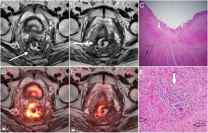
Figure 7:
PET/MRI performed in a 53-year-old man three years after surgery with a rising CEA. Axial T2 (A) demonstrates circumferential soft tissue thickening with heterogeneously mixed hyperintense and hypointense signal intensity within the surgical bed (arrow). Fused axial PET image (C) demonstrates intense FDG uptake within this circumferential soft tissue mass consistent with local recurrence. Inferiorly in the same patient, the MRI appearance is similar with semicircular T2 signal hypointensity (B, arrow), but FDG PET demonstrates an absence of hypermetabolism (D) consistent with fibrosis. At pathology, one can see both recurrent tumor (C and F, white arrows) as well as fibrosis surrounding the region of local recurrence (C and F, open arrows). The addition of FDG PET increases reader confidence and sensitivity for local recurrence.

