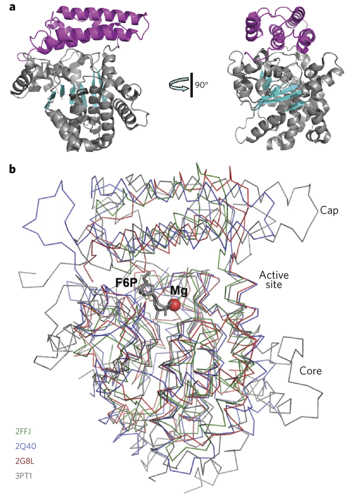
Figure 5 |. Overall structures of DUF89 protomers.
(a) The structure of YMR027W (PDB code 5BY0), shown in two orientations related by 90° rotation. The protein core domain is colored in gray (helices) and cyan (strands); the cap domain is colored in magenta. (b) Structural superimposition revealing a very similar fold of the protomers of four DUF89 structures. P. horikoshii PH1575 (PDB code 2G8L; in red), Arabidopsis At2g17340 (PDB code 2Q40; in blue), A. fulgidus AF1104 (PDB code 2FFJ; in green), and yeast YMR027W (PDB code 3PT1; in gray). A Mg2+ ion (red sphere) and a molecule of fructose 6-phosphate (F6P, gray) mark the active site cavity in the structure of YMR027W.

