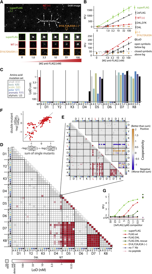
Fig 2. Binding landscape of FLAG peptide variants to M2 monoclonal antibody.
(A) A representative flow cell image shows fiducial marks and cluster-of-interest positions before the binding assay (top). Experimental images show fluorescent secondary antibody detection of binding across increasing concentrations of M2 anti-FLAG primary antibody (bottom). (B) Quantified fluorescence medians (error bars are SEM) that rise above a background threshold (grey dashed line) are extrapolated (solid) or interpolated (dashed) to estimate the limit of detection (LoD, open squares) for each FLAG variant, including WT FLAG (DYKDDDDK) and negative control (AAADDDDK) as well as superFLAG variant (DYKDEDLL), which, like 2xFLAG (DYKDDDDKGDYKDHD), gives much higher signal than WT. Two different scales are shown (top vs bottom) to accommodate the wide dynamic range of observed signals. Open symbols indicate points below background, closed symbols are above. (C) 6 amino acid mutations, each representing a physicochemical category, are represented by color (if the WT identity is the same, the alternate mutation in parenthesis is made). LoD for M2 anti-FLAG antibody binding to single mutants and (D) double mutants of the canonical (WT) peptide, DYKDDDDK, across the six amino acids at each position. (E) LoD (bottom left) and cooperativity (top right) of double mutants of the D4L base mutant (triple mutants from the WT). Cooperativity = −log(LoDDM/ LoDWT) – (−log(LoDSM1/ LoDWT) + −log(LoDSM2/ LoDWT)), where DM refers to the double mutant and SM1 and SM2 to the two corresponding single mutants. (F) Double mutant cycles. Deviations from the y=x line (grey) indicate non-additivity (cooperativity) (G) Orthogonal investigation of individual peptide variants with a fluorescence-based plate assay. Antibodies bound to variant peptides (see legend) were challenged for 2 hours with varying concentrations of 3xFLAG competitor peptide. Fluorescence values report residual bound M2 antibody.

