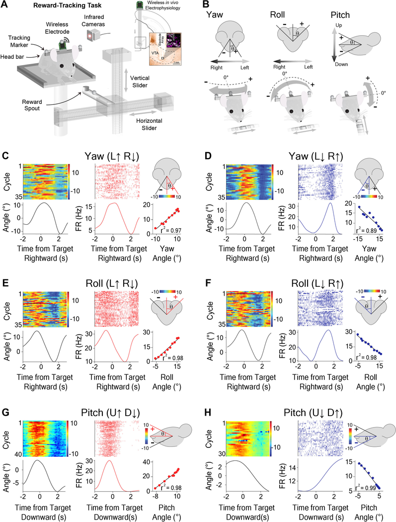
Figure 2. Opponent encoding of yaw, roll, and pitch angles by VTA GABAergic neurons.
(A) Schematic of reward-tracking task during VTA wireless in vivo electrophysiology. Mice (Wild-Type mice, n = 20) tracked a moving spout in the horizontal and vertical directions to receive a reward (diluted condensed milk). Infrared cameras captured kinematic data from markers on the head of the animal with respect to reward target. Inset shows schematic of electrode placement into the VTA and representative histological section in the vicinity of DAT-positive cells.
(B) Schematic representation and sign conventions for yaw (left), roll (middle), and pitch (right) rotational kinematics. Arrows show direction of head movement around each axis. Zero angle is when the animal has his head straight forward.
(C-H) Individual neurons represent direction-specific angles along orthogonal axes of rotation. Peri-event heat maps of head angle (left), peri-event raster plots of VTA neural activity (middle) with respect to reward target, and a correlation graph (right bottom) with a schematic illustration demonstrating head angle direction for which the firing rate increases (right top).
(C) Yaw angle and Yaw (L↑ R↓) neuron during horizontal tracking (Pearson Correlation (PC), r2 = 0.97, p < 0.0001).
(D) Yaw angle and Yaw (L↓ R↑) neuron during horizontal tracking (PC, r2 = 0.89, p < 0.0001).
(E) Roll angle and Roll (L↑ R↓) neuron during horizontal tracking (PC, r2 = 0.98, p < 0.0001).
(F) Roll angle and Roll (L↓ R↑) neuron during horizontal tracking (PC, r2 = 0.98, p < 0.0001).
(G) Pitch angle and Pitch (U↑ D↓) neuron during vertical tracking (PC, r2 = 0.98, p < 0.0001).
(H) Pitch angle and Pitch (U↓ D↑) neuron during vertical tracking (PC, r2 = 0.99, p < 0.0001). See also Figures S1–S3 and S7, Videos S1–S3.

