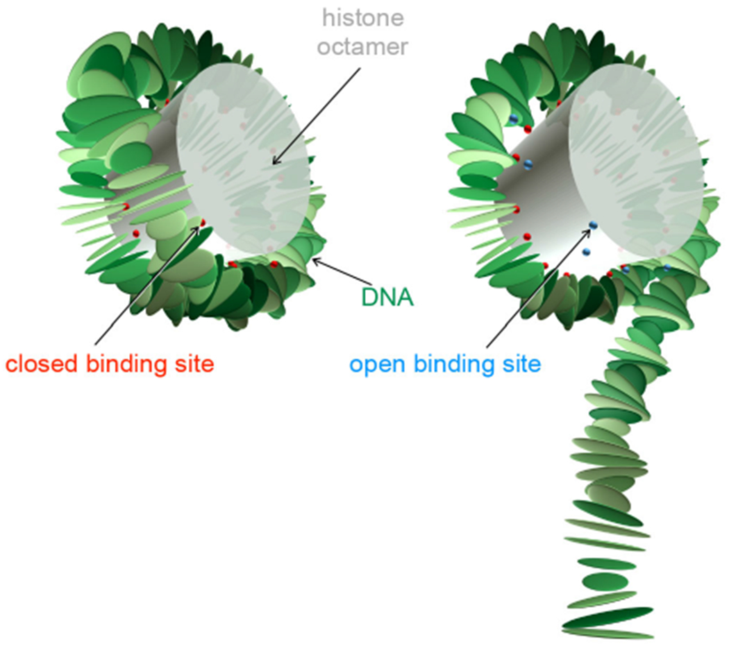
Fig. 1.

Nucleosome model with the fully wrapped complex on the left and a partially unwrapped complex on the right. Each rigid plate represents a bp, the locations of the constraints (corresponding to bound phosphates) are shown by beads, two per binding site. Red beads represent closed sites and lue beads open sites. The cylinder is a rough representation of the protein core but is not simulated explicitly (except through the binding sites).
