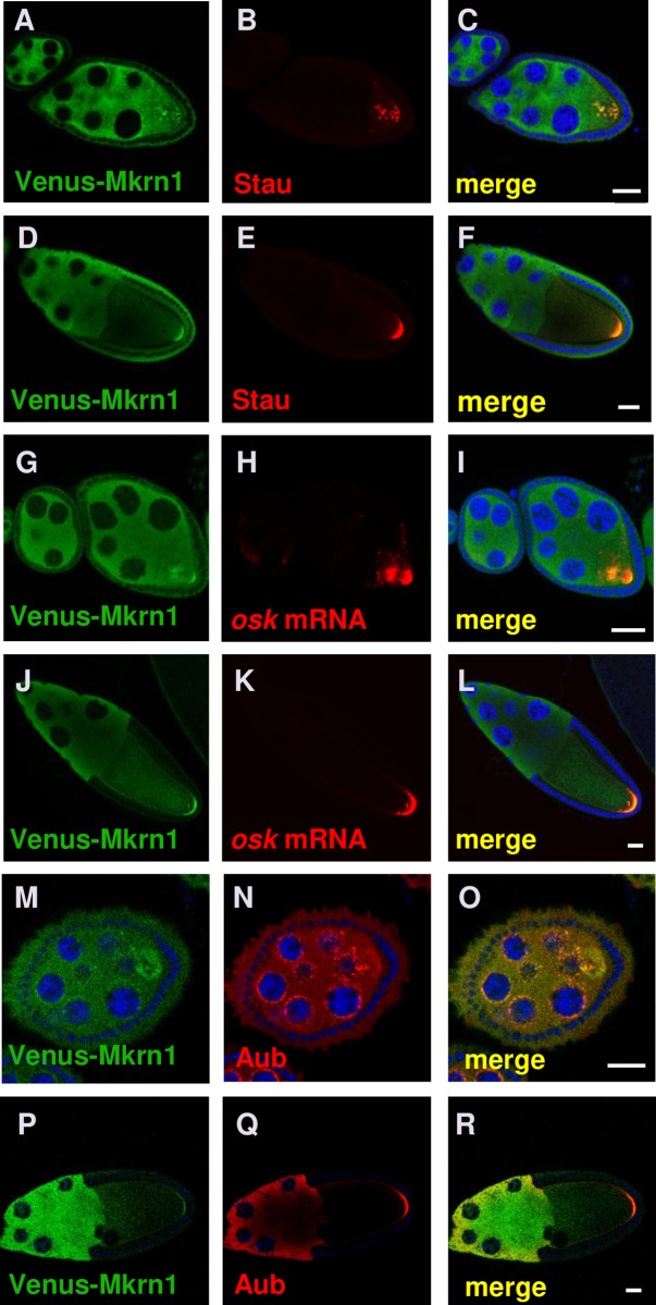
Fig 2. Mkrn1 accumulates in pole plasm.
(A-C) The three panels show the same egg chambers stained for (A) Venus-Mkrn1, (B) Stau, and a (C) merged image. Scale bars, 25 μm. Venus-Mkrn1 expression was driven by nos>Gal4. Colocalization of Venus-Mkrn1 and Stau can be observed in particles that have not yet accumulated at the posterior of the early stage 8 oocyte. (D-F) The three panels show the same stage 10 egg chamber stained for (D) Venus-Mkrn1, (E) Stau and (F) a merged image. Scale bars, 25 μm. There is extensive colocalization of Venus-Mkrn1 and Stau in the posterior pole plasm of the oocyte. (G-I) The three panels show the same egg chambers stained for (G) Venus-Mkrn1, (H) osk mRNA, and (I) a merged image. Scale bars, 25 μm. Colocalization of Venus-Mkrn1 and osk can be observed in an early stage 8 oocyte where osk has not yet fully localized at the posterior of the oocyte. (J-L) The three panels show the same stage 10 egg chamber stained for (J) Venus-Mkrn1, (K) osk mRNA and (L) a merged image. Scale bars, 25 μm. There is extensive colocalization of Venus-Mkrn1 and osk mRNA in the posterior pole plasm of the oocyte. (M-O) The three panels show the same egg chambers stained for (M) Venus-Mkrn1, (N) Aub, and a (O) merged image. Scale bars, 5 μm. Colocalization of Venus-Mkrn1 and Aub can be observed at the nuage surrounding the nurse cell nuclei (P-R). The three panels show the same egg chambers stained for (P) Venus-Mkrn1, (Q) Aub, and a (R) merged image. Scale bars, 20 μm. There is extensive colocalization of Venus-Mkrn1 and Aub in the posterior pole plasm of the oocyte.

