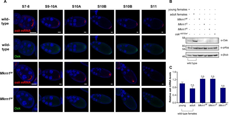
Fig 4. Translation of osk mRNA is impaired in Mkrn1W ovaries.
(A) Fluorescent in situ hybridization for osk mRNA (red) with co-immunostaining for Osk protein (green) in wild-type and Mkrn1W egg chambers. For each genotype and in each column the top and bottom images are of the same egg chamber. In wild-type oocytes posterior accumulation of osk mRNA and Osk protein is robust and stable from stage 9 onward. In Mkrn1W oocytes accumulation of osk mRNA resembles the wild-type pattern through stage 10A but is not maintained, while Osk protein is rarely detectable at the oocyte posterior at any stage. Scale bars, 25 μm. (B) Western blot analysis from ovary lysates of various genotypes stained for Osk, pAbp and β-tubulin. Osk protein levels are greatly reduced in all Mkrn1 mutant alleles. 1 day-old young females have not yet completed oogenesis and were used as a control for Mkrn1S and Mkrn1N ovaries which also lack late-stage egg chambers, where Osk is most abundant. (C) RT-qPCR experiments measuring ovarian osk mRNA levels (normalized to RpL15 mRNA) in the same genotypes as (B). mRNA levels of ovaries from adult females were compared to Mkrn1W ovaries. For Mkrn1S and Mkrn1N ovaries, mRNA levels of 1 day-old young ovaries was used as normalization. Error bars depict Stdev, n = 2.

