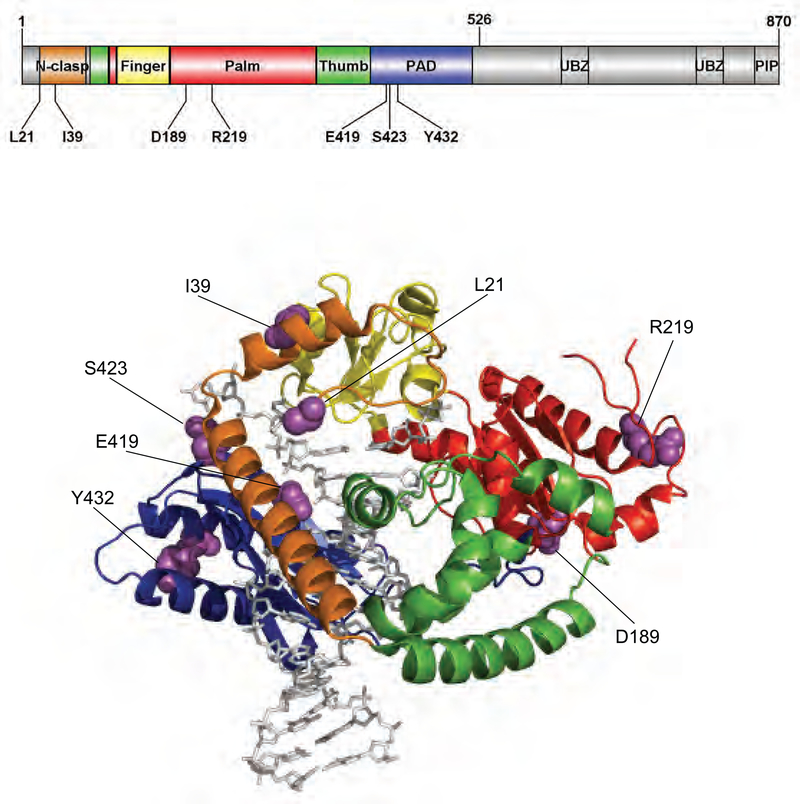
Figure 5. Locations of genetic pol κ variations.
Structure of human pol κ21−517 (PDB code, 2OH2) bound to primer/template DNA and incoming nucleotide is shown using Pymol. Pol κ21−517 is shown in cartoon ribbons, and the primer/template DNA and dNTP are shown in gray sticks. The N-clasp, finger, palm, thumb, and PAD domains are colored orange, yellow, red, green, and blue, respectively. The amino acid residues (in purple spheres) of genetic pol κ variants are indicated. The structural domains of pol κ are shown in the upper schematic diagram using DOG (version 2.0),54 where locations of amino acids related to eight studied variations are indicated.

