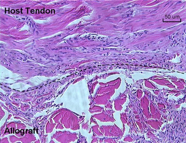
Fig. 11.
The junction of the host flexor digitorum profundus tendon and the carbodiimide-derivatized hyaluronic acid and gelatin-treated allograft at the proximal repair site. The top of the dashed line is the host tendon. The bottom of the dashed line is the allograft. Tenocytes were observed in the allografts (H & E, ×200).

