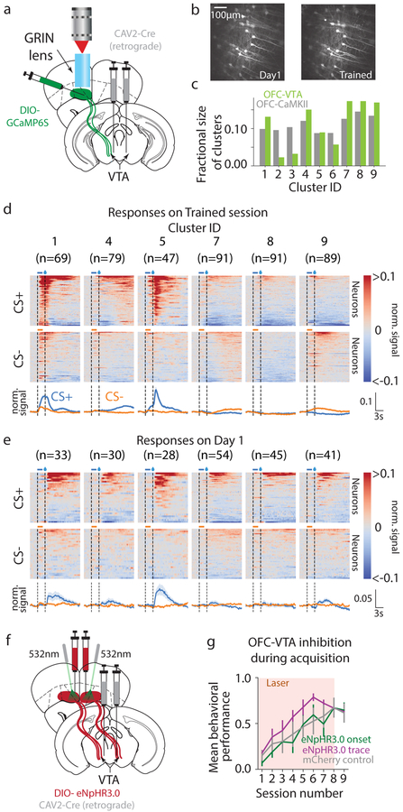
Fig. 3. OFC-VTA neurons convey information selective to cue-reward associations.
a. Schematic of imaging experiment to record from vmOFC neurons projecting to VTA (OFC-VTA). b. Example activity projection maps of OFC-VTA cells on Day 1 and Trained. c. Fraction of neurons per cluster in OFC-VTA compared to OFC-CaMKII population. We restricted further analysis to those clusters in which we could identify at least 2 cells on average per animal in the imaging plane tracked over learning, which also excluded cluster 6 d. PSTHs during Trained session showing responses of clusters. e. PSTHs of neurons in these clusters on Day 1, showing the absence of cue onset responses. f. Schematic of optogenetic experiment to target OFC-VTA neurons. g. Behavioral acquisition of reward seeking to CS+ during inhibition of OFC-VTA neurons showed no effect during cue onset or trace periods. Reduced behavioral performance of all groups compared to OFC-CaMKII group is likely due to a difference in age (Methods). Measure of center is the mean and error bars represent standard error of the mean.

