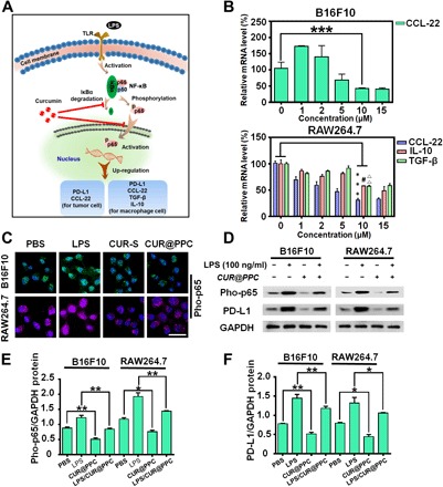
Fig. 3. CUR released from CUR@PPC suppressed cytokines (CCL-22, TGF-β, and IL-10) secretion and PD-L1 expression in tumor.

(A) Schematic illustration of CUR to suppress cytokines (CCL-22, TGF-β, and IL-10) secretion and PD-L1 expression via inhibiting the NF-κB pathway of tumor cells and macrophages. (B) Inhibited expressions of the CCL-22 gene in B16F10 cells and the CCL-22, IL-10, and TGF-β genes in RAW264.7 cells by CUR@PPC determined by real-time reverse transcription PCR (n = 3; means ± SD; ***P < 0.001, #P < 0.05, ΔΔP < 0.01). (C) CLSM images showed that CUR@PPC significantly inhibits the NF-κB pathway of B16F10 and RAW264.7 cells. Pho-p65 was labeled with Alexa Fluor 488 (green fluorescence) in B16F10 cells or Alexa Fluor 647 (purple fluorescence) in RAW264.7 cells (concentration of CUR@PPC, 10 μM). Scale bar, 25 μm. (D) Western blot assay showed that the NF-κB pathway and PD-L1 expression in B16F10 cells and RAW264.7 cells were inhibited by CUR@PPC (concentration of CUR@PPC, 10 μM). GAPDH, glyceraldehyde phosphate dehydrogenase. Protein expression levels of PD-L1 (E) and pho-p65 (F) quantified from Western blot. (n = 3; means ± SD; *P < 0.05, **P < 0.01). Statistical analyses were performed using analysis of variance (ANOVA) with Tukey’s test.
