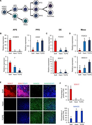
Fig. 2. Soft substrate induces endodermal lineage commitment.

(A) A schematic drawing of the differentiation of hPSCs into liver and pancreatic cells. Meso, mesoderm; AFG, anterior foregut; PFG, posterior foregut; MHG, midgut/hindgut. Day 3 mRNA expression of hPSC genes involved in (B) APS/PPS, (C) DE, and (D) mesoderm differentiations. The results are presented as means ± SD of triplicates. One-way analysis of variance (ANOVA; n = 3 independent experiments). (E) Immunofluorescent staining and (F) quantification of SOX17 and NANOG in hPSCs grown on substrates with different stiffnesses. The results are presented as means ± SD of triplicates. One-way ANOVA (n = 3 independent experiments). Different letters indicate significant differences, and the same letters indicate no significant difference.
