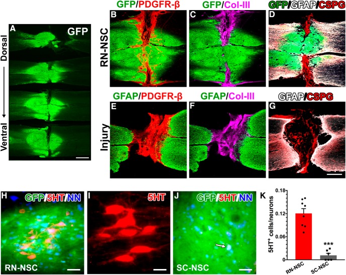
Figure 2.
Grafted E14 BS-NSCs partially integrate into the spinal cord. A, Surviving GFP+ BS-NSC implants are shown in a series of longitudinal spinal cord sections from the dorsal to ventral. Grafts form a bridge in the middle part and connect the rostral and caudal cords. B–G, Immunostaining revealed that (B) numerous PDGFR-β+ fibroblasts are present in the portions of the lesion devoid of GFP+ tissue. Col-III (C) and CSPG (D) are heavily deposited in this region. There is a little expression of GFAP at the interface of host/graft. In injury controls (E–G), however, a dense GFAP+ glial scarring is often present at the stumps of spinal cord. Intense Col-III and CSPG are stained into the lesion site of spinal cord. H–K, Abundant serotonergic neurons grow in RN-NSC grafts (H,I), whereas very few are detected (arrow) in SC-NSC grafts (J). These neurons express NeuN, indicating that they have developed mature. K, Cell quantification demonstrated that the percentage of serotonergic neurons in RN-NSC grafts is significantly higher (unpaired t test, ***p < 0.001) than those in SC-NSC grafts (n = 6 or 8 per group). Scale bars: A, 2 mm; G, 0.5 mm; H, J, 50 μm; I, 20 μm.

