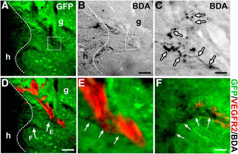
Figure 5.

Descending vasomotor pathways regenerate into NSC grafts. A–C, BDA injection into the RVLM labels regenerated supraspinal axon terminals within the GFP+ RN-NSC-grafted region in the spinal cord (n = 5). Dotted lines indicate the interface of host (h)/graft (g). Labeled descending axon terminals (arrows) mainly emerge in the rostral part of GFP+ grafts. C, Higher magnification of the boxed region in A and B. D–F, Costaining of BDA and immunofluorescent labeling of VEGFR-2 disclose that penetrated axon terminals often apposite along nascent blood vessels in GFP+ implants. Scale bars: B, D, 100 μm; C, 12.5 μm; F, 30 μm.
