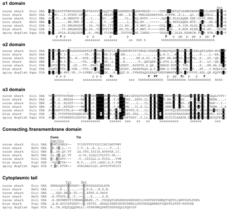
Figure 1.
Multiple amino acid alignment of the four distinctive MHC class I lineages in cartilaginous fish, UAA, UBA, UCA and the new lineage UDA. GenBank accession numbers are listed in Supplemental Table I. Dots indicate amino acids identical to Gici UAA, and dashes indicate gaps, respectively. Highly conserved residues in all class I proteins are shaded black (16, 28), s and h indicate the β-strands and α-helices, and the line connecting the two Cys in the α2 domain indicates the class I canonical disulfide bridge. P marks the invariant residues that bind to the N- and C-termini of the bound peptide in the classical class I molecules and p indicates the other 28 peptide binding residues. The Asn marks the Asparagine-linked glycosylation site, Asp/Glu indicates the typical Aspartic acid and Glutamic acid residues found in the connecting piece (Conn, light shade), and Tyr and Ser mark the conserved positions of Tyr and Ser in the cytoplasmic tail (also in light shade) of classical class I molecules.

