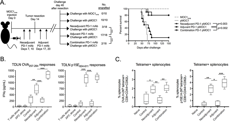
Figure 3 – Mice rejected engraftment with tumor cells lacking OVA following neoadjuvant PD-1 mAb.
A, MOC1ova tumors were established in WT B6 mice and completely resected at day 14. Tumor-bearing mice were given neoadjuvant (days 7 and 10), adjuvant (days 17 and 20) or combination PD-1 mAb. 40 days after resection, untreated mice were challenged with MOC1ova or pMOC1 cells as controls, and treated mice were challenged with pMOC1 cells and followed for engraftment. Tumor engraftment rates are shown on the left, and survival to 150 days after challenge is shown as a Kaplan-Meier curve on the right. Cumulative data from two independent experiments.
B and C, MOC1ova tumor-bearing mice were treated neoadjuvant, adjuvant or combination PD-1 mAb as in A. B, 40 days after resection, T cells were sorted form tumor draining lymph nodes (TDLN) and assessed for IFNγ production upon exposure to antigenic peptides by ELISA. C, 40 days after resection, splenic T cells were assessed for tetramer and memory marker positivity by flow cytometry (n = 5 independent tumors each condition).
Unless stated otherwise, all data shown is representative data from one of at least two independent experiments.
*, p < 0.05; **, p < 0.01; ***, p < 0.001, student’s t-test

