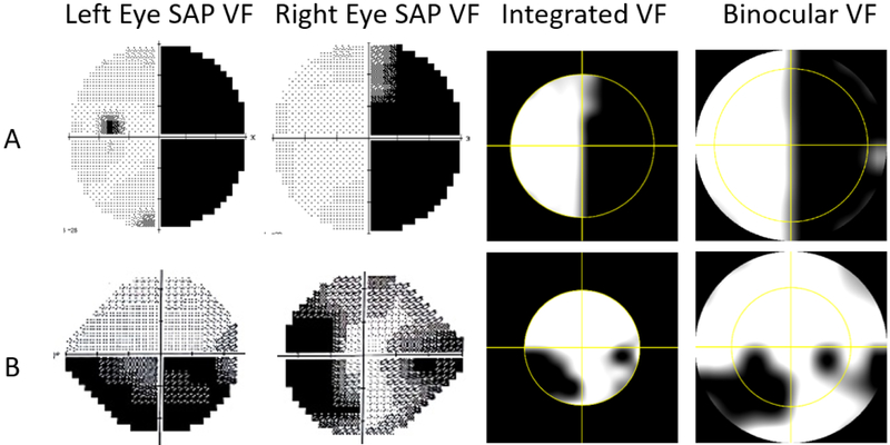
Figure 4:
Binocular visual field (VF) measurements for two patients A) Hemianopia patient tested with 30–2 monocular Humphrey standard automated perimetry (SAP). B) Glaucoma patient tested with 24–2 Humphrey SAP. First column: left eyes monocular SAP. Second column: Right eyes Monocular SAP. Third column: binocular Integrated VF (IVF) constructed by merging the two monocular fields based on the maximum sensitivity model. Fourth column: Direct binocular VF measurement with the digital spectacles.

