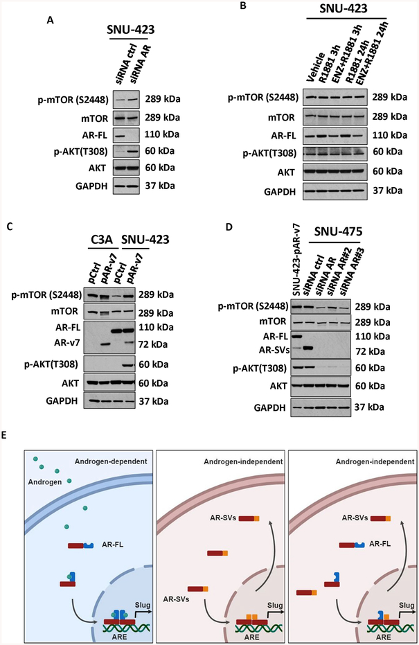
Figure 6. Androgen-dependent and Androgen-independent signaling in HCC.
(a) SNU-423 were transfected with siRNA control or siRNA against AR (as in 5F). Relative to siControl cells, siRNA AR-transfected cells demonstrated upregulation of both phosphorylated mTOR and AKT with no change in total mTOR and AKT. (b) AR expressing HCC cells SNU-423 were treated with vehicle, 1 nM R1881 or 10uM enzalutamide with 1 nM R1881 for 3 and 24 hours. Relative to vehicle-treated cells, no change in protein expression of total or phosphorylated mTOR or AKT was apparent following treatment. (c) AR-negative, C3A, and AR-expressing SNU-423 HCC cells were transiently transfected with either 10 μg AR-v7 expressing plasmid (pAR-v7) or control (pcw107, pControl). Relative to pControl, AR-v7-overexpressing cells showed an upregulation of phosphorylated mTOR and AKT with no change in the total levels of mTOR and AKT. AR protein levels in C3A also shown in Figure 3G (d) AR-Sv expressing HCC cells SNU-475 were transfected with control siRNA (siControl) or 3 different siRNA against AR and compared to AR-v7 transfected SNU-423 cells. Relative to siControl, siRNA AR-transfected SNU-475 cells showed a downregulation of both phosphorylated mTOR and AKT with no change in the protein levels of total mTOR and AKT. (e) Graphical depiction of potential AR signaling to modulate EMT in HCC, androgen-dependent AR-FL homodimers (left), androgen-independent AR-Svs homodimers (middle) and androgen-independent AR-FL and AR-Svs heterodimers (right).

