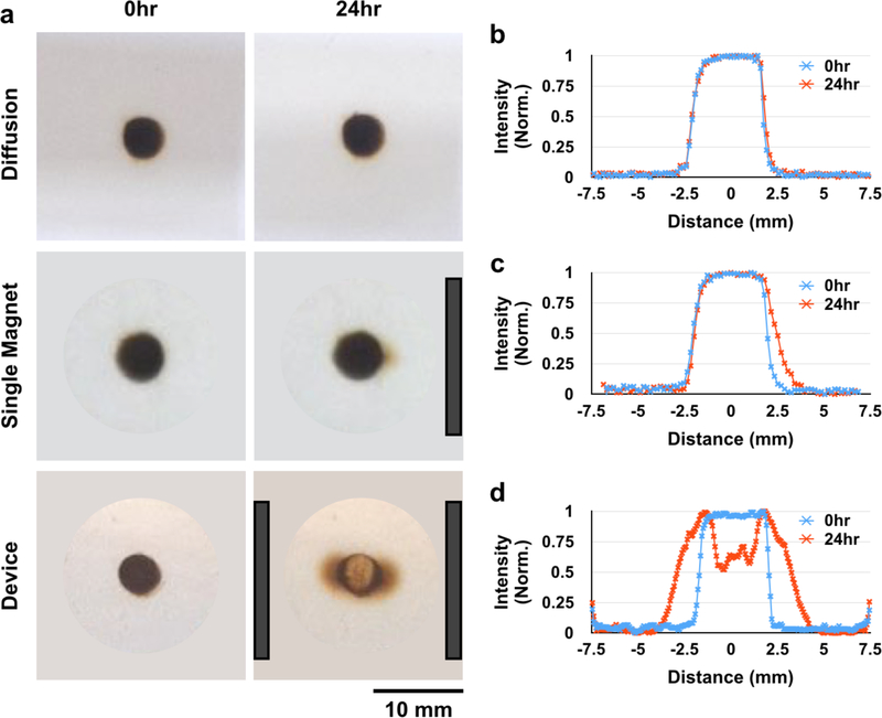
Figure 6.
The magnetic device radially disperses SPION micelles through a 0.4% agarose tissue phantom. There is minimal diffusion of SPION micelles through the tissue phantom over 24 hours (a: top, b). When placed next to a single magnet, SPION micelles move slightly through the gel toward the direction of the magnet (a: middle, c). However, in the magnetic device, particles disperse radially away from the zero point of the device (a: bottom, d). Magnet positions are indicated by dark gray bars.

