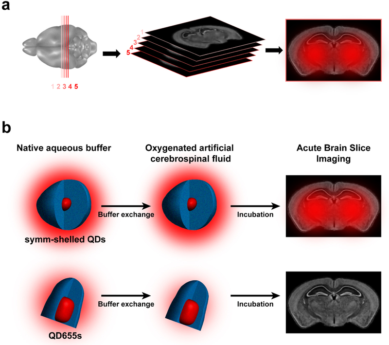
Fig. 4.
(a) Acute brain slice imaging with CdSe/CdS QDs. Mouse brain slices (1–5 300 μm slices) are cut by vibratome and incubated with ligand-conjugated QDs prior to imaging. (b) Schemes outlining buffer exchange of QDs (drawn to scale) into brain slice media (oxygenated artificial cerebrospinal fluid, aCSF) for symmetrically shelled QDs and commercial QD655s. The schemes illustrate comparison of symmetrically shelled QD and QD655 performance in oxygenated aCSF and their photoluminescence fate in tissue specimens. The auras surrounding QD structures illustrate relative photoluminescence and the fate of diminished performance of QD655 in slice media. Whole brain slice representations are provided by the Allen Institute.44

