FIG 2.
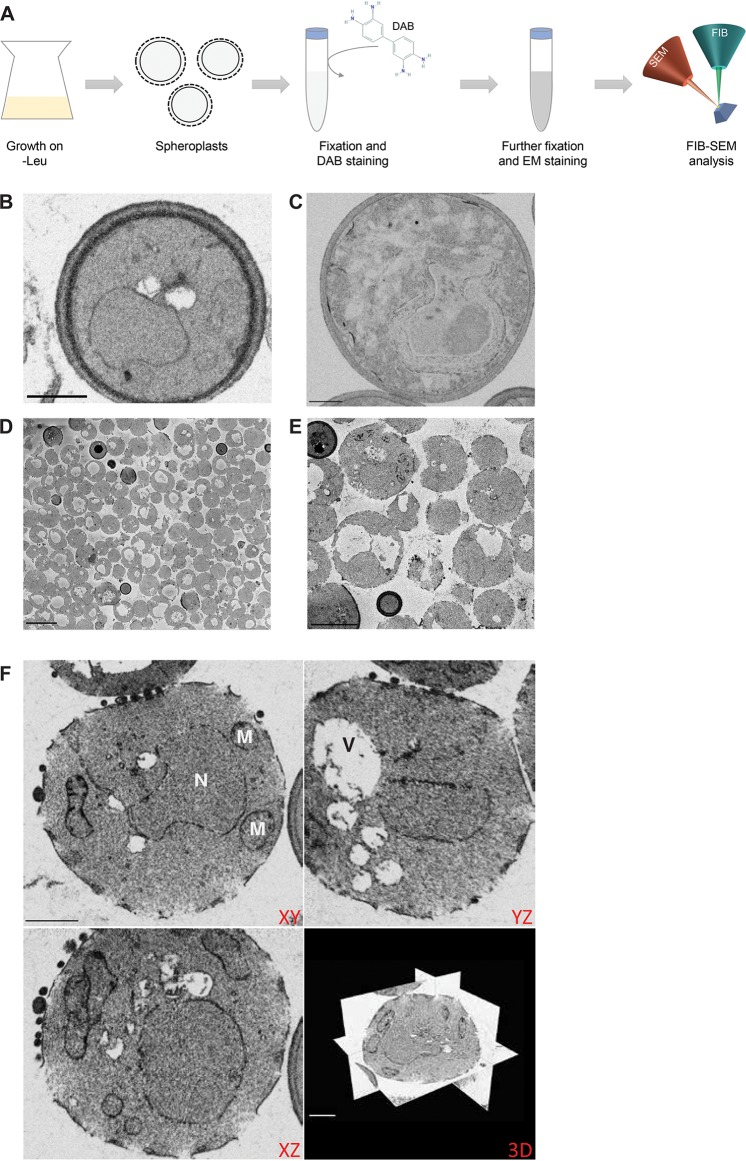
Spheroplast formation does not interfere with FIB-SEM sample preparation. (A) The workflow of the experiment is presented, where cells are grown overnight in −Leu medium and subsequently subjected to spheroplast formation. The spheroplasts next are fixed, incubated with DAB (structure retrieved from the PubChem Database, CID = 7071; https://pubchem.ncbi.nlm.nih.gov/compound/7071), and further prepared for FIB-SEM analysis. (B and C) FIB-SEM images of nonspheroplast control cells expressing Erg11-V5 without the APEX2 tag (B) and nonspheroplast cells expressing Erg11-V5-APEX2 (C) show low electron-dense signal in both samples (scale bars, 1 μm). (D and E) Representative images of SBF-SEM data of control cells after spheroplast formation, showing that spheroplast formation was successful for most cells (D) and organelle ultrastructure was preserved (E) (scale bars, 5 μm). (F) Orthogonal views of FIB-SEM data of the same sample of control cells confirm the preservation of organelle ultrastructure and cell shape (scale bars, 1 μm). N, nucleus; M, mitochondria; V, vacuole.

