FIG 4.
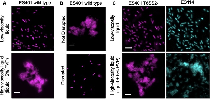
V. fischeri forms aggregates in high-viscosity, liquid medium. Fluorescence microscopy images of GFP-tagged ES114 (cyan) or RFP-tagged wild-type or vasA_2 mutant (T6SS2 mutant) ES401 (magenta) incubated in the specified medium type for 12 h. (A) The ES401 wild type was incubated in either low-viscosity liquid or high-viscosity liquid. (B) The ES401 wild type was incubated in high-viscosity liquid and either spotted directly onto a glass slide (Not Disrupted) or mixed prior to spotting (Disrupted). (C) The ES401 vasA_2 mutant and ES114 were incubated in either low-viscosity liquid or high-viscosity liquid. For all fluorescence microscopy images, each experiment was performed twice with two biological replicates and five fields of view. One representative image is shown. Bars = 5μm.

