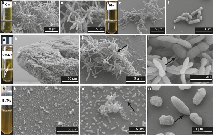
FIG 3.
Scanning electron micrographs of the cultures at 3 to 7 days of growth. (a, d, g, and k) Representative Balch tubes of cultures of C. minuta (Cm), M. smithii (Ms), C. minuta and M. smithii (Cm/Ms), and B. thetaiotaomicron and M. smithii (Bt/Ms) after 7 days of growth; (b and c) scanning electron micrographs (SEMs) of monocultures of C. minuta at 5 days of growth; (e and f) SEMs of monocultures of M. smithii at 5 days of growth; (h to j) SEMs of cocultures of C. minuta and M. smithii at 7, 5, and 2 days of growth, respectively; (l to n) SEMs of cocultures of B. thetaiotaomicron and M. smithii at 7 days of growth. The floc formed by C. minuta and M. smithii is indicated with a white arrow in panel g; other arrows indicate M. smithii cells. Metal bars in panels a, d, and g are from the tube rack.

