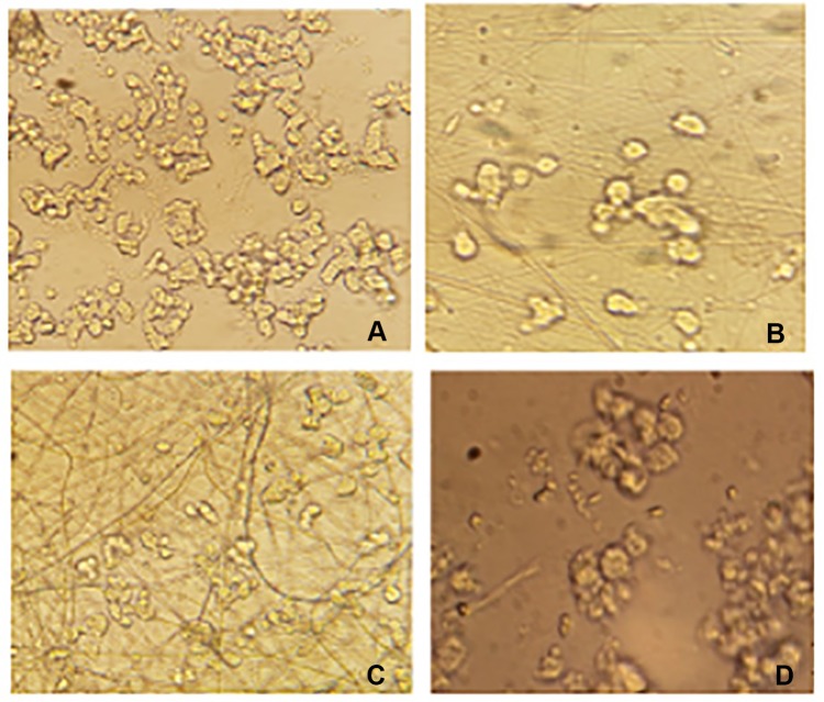
Figure 7.
The morphology of PC12 cells cultured on the nanofiber samples (A) PAN-kefiran 5% after 6 days of cell seeding (magnifying×400). (B) PAN-kefiran 10% after 6 days (magnifying×400). (C) PAN nanofiber sample after 6 days (magnifying×400). (D) The control sample (without any treatment) after 6 days (magnifying×400).

