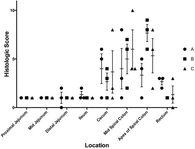
Figure 3.

Histologic scores from samples collected at necropsy on day 7 following inoculation with three separate isolates of S. 4,[5],12:i:- (A, B, C) in animal study #1. These scores depict the average histologic lesions, as determined by the ulceration, neutrophil infiltration, and crypt elongation and abscessation, and submucosal inflammation, at the time of necropsy on DPI 7. The mean of each isolate-tissue location combination is represented by the horizontal bar with the standard error represented by the vertical line.
