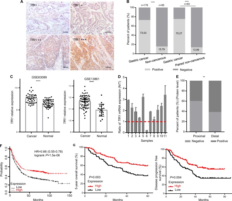
Fig. 1. TfR1 protein expression in GC patients reversely correlated with poor prognosis.
a Different staining scores with M-HFn nanoparticles detecting TfR1 in GC tissues by IHC, scale bars: 50 µm. b Expression level of TfR1 protein in GC and their (or matched) adjacent noncancerous tissues. c TfR1 mRNA expression was significantly upregulated in GC tissues compared with adjacent normal mucosa in GES63089 and 13861 from GEO datasheets, respectively. d Ratio (T/N) of TfR1 mRNA expression in 11 paired primary GC patients, which was determined by qPCR (lower panel). Their expression levels were normalized by an internal control (GAPDH). e IHC staining that indicated high TfR1 protein expression was significantly associated with location of GC. f Kaplan–Meier survival analysis of OS obtained from public gene expression datasets. g Kaplan–Meier analysis of 5-year survival with low vs. high TfR1 protein expression status (left); Kaplan–Meier survival analysis of DFS with low vs. high TfR1 protein expression status (right). *P < 0.05; **P < 0.01; ****P < 0.0001.

