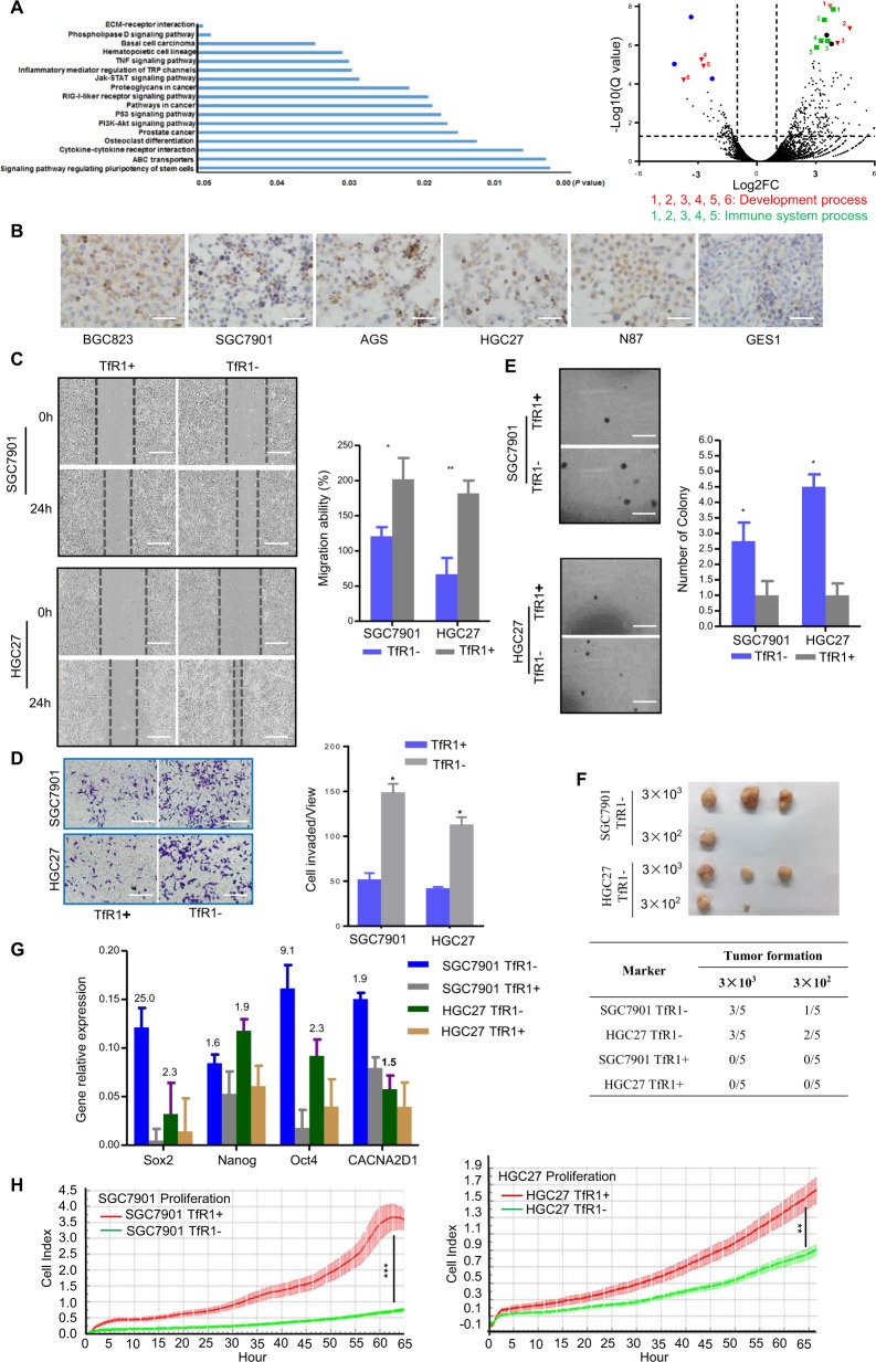
Fig. 3. GC cells with the absence of TfR1 possess tumor-initiating like properties through in vitro and in vivo assays.
a RNA-seq profiles for sorted TfR1-negative and -positive cells were analyzed. Significant signaling pathway (left panel) and volcano plot illustrated the differentially expressed genes between TfR1-negative and -positive cells (right panel, fold change > 2.0 or <2.0; Q value < 0.05). Blue, green, and red colors indicated various genes belonging to different groups of cell processes. b TfR1 was overexpressed in six GC cells (BGC823, SGC7901, AGS, HGC27, N87, and GES1). c–e Absence of TfR1 promoted cell migration, invasion, and colonogenicity by wound-healing assay, Boyden chamber invasion assay, and colony formation assay. Scale bar: 100 µm. f Analysis of TfR1 sorted ± cell tumorigenity following transplantation with different numbers of cells into NOD/SCID mice. g Sox2, Nanog, Oct4, and CACNA2D1 mRNA relative expression was determined by qPCR. Their expression levels were normalized by an internal control (GAPDH). h TfR1-positive sorted cells showed obviously cell proliferation ability detected by real-time RTCA instrument. *P < 0.05; **P < 0.01, ***P < 0.001.

