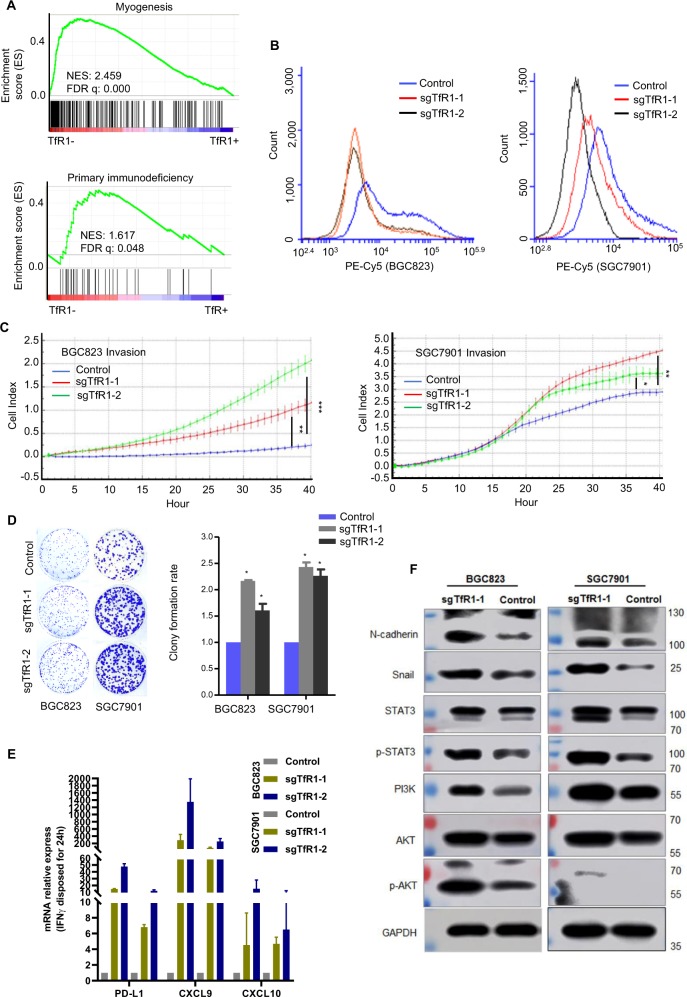
Fig. 5. TfR1-knockout cells displayed malignant properties and responded to IFN-γ treatment.
a GSEA plot based on RNA-seq (upper panel) or TCGA data (lower panel) indicated TfR1-negative cells correlated with developmental and immune deficiency. b TfR1-knockout cells were identified by flow cytometry analysis. c TfR1-knockout GC cells showed high invasion ability evaluated by RTCA real-time analysis instrument. d Knockout of TfR1 increased cell colonogenicity. e PD-L1, CXCL9, and CXCL10 mRNA level was enhanced in TfR1-knockout cells stimulated with IFN-γ (100 ng/ml) for 24 h. f The correlation of EMT- related markers, STAT3/AKT signaling with TfR1, were detected by Western blot. NES normalized enrichment score. *P < 0.05; **P < 0.01; ***P < 0.001.

