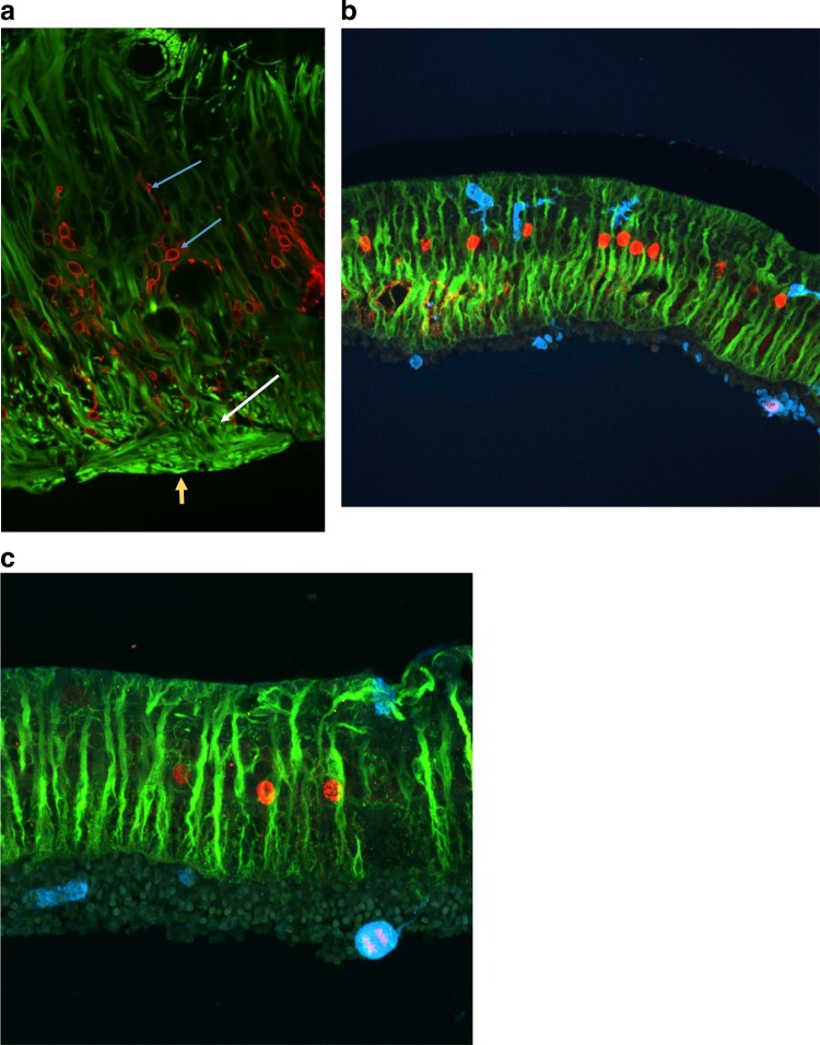
Fig. 1.
a Confocal microscopy of human PVR retinectomy specimen. Glial intermediate filament stain (GFAP) green and rhodopsin stain (red). Note marked upregulation of glial intermediate filament expression throughout retina. The photoreceptor inner and outer segments have been lost and glial cell processes have extended through the outer limiting membrane (white arrow) to form a confluent subretinal glial scar (yellow arrow). Rhodopsin has been redistributed to rod cell bodies and rods have extended neurite processes towards the inner retina (blue arrows). b, c Experimental retinal detachment (3 day, rabbit). Anti-Vimentin (green) labels Muller cells. There is an increase in labelling within Muller cells after detachment. Anti-BrdU (red) labels dividing cells. Isolectin B4 (blue) labels microglia. b (Control, untreated) proliferating glial (Muller) cells are demonstrated by the red stain (anti BrdU) in the inner nuclear layer. There is an increase in dividing Muller cells (in the INL) and microglia (one shown in the OS layer). c Treated with 2 mg triamcinolone on detachment induction and at 24 h note the marked reduction in glial cell proliferation (from a mean of 21 cells/mm retina to 7 cells/mm retina)

