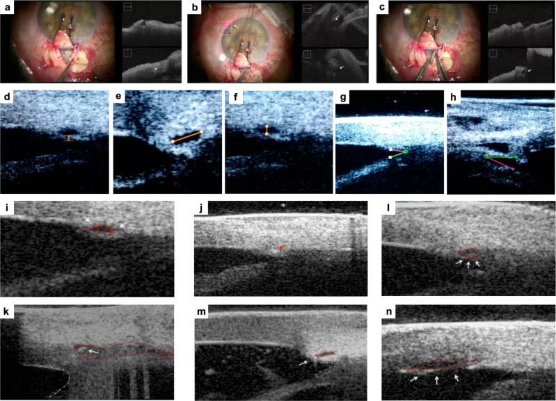
Fig. 1.
Picture a Microscope image (left picture) shows trabeculo-descemetic window after dissection of the deep scleral flap. iOCT image (right picture) shows SC before dilating the ostium of SC using the iTrack microcatheter (white arrow). Picture b Microscope image (left picture) shows the progress of microcatheter into SC. Red light indicates the position of the microcatheter tip in the SC. iOCT image (right picture) shows the microcatheter in the SC (white arrows) Picture c Microscope imaging shows the final step of retraction of the microcatheter with simultaneous injection of Healon GV into the SC. iOCT image (right picture) shows the enlarged SC (white arrow) Picture d Intraoperative UBM image: TM thickness measurement after SC viscodilation. Picture e, f Intraoperative UBM image: horizontal and vertical diameter of SC after viscodilation. Picture g, h Intraoperative UBM image: AC angle before (g) and after surgery (h) Picture i Preoperative iUBM shows SC that appears as an anechogenic oval-shaped space (red dashed line), and TM (white arrow) Picture j iUBM shows the microcatheter in the SC that appears hyperechogenic in comparison to the surrouding tissue (red arrow) Picture l: iUBM shows enlarged SC after viscodilation (white arrows, red dashed line) Picture k: iUBM shows SC with suture before tensioning suture (white arrow), deep scleral dissection and filtering window (red dashed line). AC angle appears with a concave and rounded profile (white dashed line) Picture m iUBM shows SC and suture after tensioning suture (white arrow). SC appears elongated (red dashed line) and AC angle becomes acute (white dashed line) Picture n iUBM shows Descemet’s membrane detachment after viscodilation (white arrows and red dashed line)

