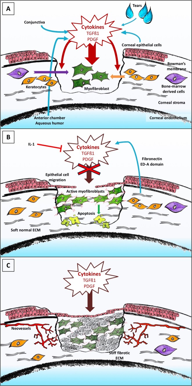
Fig. 2.
Schematic diagram of the corneal wound healing process. a Implementation of the healing process. Corneal injury involves rupture of the corneal epithelial layer and of the Bowman’s membrane. The anterior stroma therefore is highly exposed to cytokines and others growth factors, particularly transforming growth factor-β1 (TGFß1) and platelet-derived growth factor (PDGF), from corneal epithelial cells, aqueous humour, conjunctiva and tears. Under the influence of active TGFß1, keratocytes of the anterior stroma and/or cells derived from bone marrow, which normally are in a quiescent state, transdifferentiate into myofibroblasts, proliferate and then spread to the site of the lesion by centripetal migration. b Physiological wound healing. Epithelial cells migrate and epithelial proliferation appears. Myofibroblasts synthesize new extracellular matrix (ECM). In the absence of pathological phenomena, myofibroblasts disappear by interleukin (IL)-1-induced apoptosis and tissue transparency is maintained. c Pathological conditions. Myofibroblasts live on and continue to secrete excessive amounts of aberrant ECM proteins. This situation leads to fibrosis and the clinical onset of corneal opacification. This chronic corneal injury subsequently induces corneal neoangiogenesis

