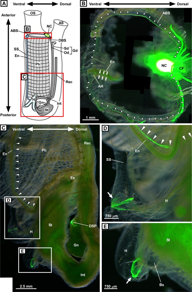
Figure 1.

Overview of the distribution of PC2 promoter-driven Kaede-positive cells in transgenic Ciona. (A) Schematic illustration of an adult Ciona. The key anatomic parts of an adult Ciona are indicated by different fill patterns. (B) Kaede-positive cells in the framed region shown in (A). The anterior end of the pharynx viewed from above is shown. Arrowheads indicate Kaede-positive cells distributed along the anterior pharyngeal blood sinus located at the anterior end of the pharynx. Arrows indicate Kaede-positive cells in the endostyle. The oral siphon was removed to make the blood sinus more visible. Thirty-one images were merged to construct a composite image. The neural complex has a very strong Kaede-positive signal that was described in the previous study18. Note that the ciliated funnel is not Kaede-positive, but Kaede signals from the neural complex and nerve fibers are shown through the ciliated funnel. (C) Kaede-positive cells in the posterior half of the transgenic Ciona framed in (A). Peripheral organs, including the endostyle, pharynx, heart, and stomach are shown. Arrowheads indicate Kaede-positive cells in the endostyle. Kaede-positive signals were also observed in the heart, stomach, and dorsal strand plexus. Kaede-positive signals in the pharynx were relatively weak and not visible in this image. The tunic, body-wall muscle, and part of the pharynx were removed to make the peripheral organs more visible. Seven images were merged to construct a composite image. (D) Magnified image of the framed region in (C). The junction between the heart and blood sinus adjacent to the endostyle is shown. The left outlet of the heart is surrounded by Kaede-positive cells (arrow). Kaede-positive cells in the endostyle are indicated by arrowheads. Two images were merged to construct a composite image. (E) Magnified image of the framed region in (C). The junction between the heart and blood sinus connecting to the stomach is shown. The right outlet of the heart is surrounded by Kaede-positive cells. These images were obtained under a fluorescence stereomicroscope as superimposed images. ABS, anterior pharyngeal blood sinus, AH, anterior hood; AS, atrial siphon; BS, blood sinus; CF, ciliated funnel; DBS, dorsal blood sinus; DSP, dorsal strand plexus; En, endostyle; Es, esophagus; Gd, gonoduct; Gn, gonad; H, heart; Int, intestine; NC, neural complex; Od; oviduct, OS, oral siphon; Ph, pharynx; Rec, rectum; Sd, spermiduct; SS, subendostylar sinus; St, stomach.
