Figure 2.
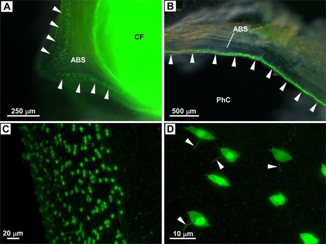
Distribution of PC2 promoter-driven Kaede-positive cells in the blood sinus. (A) Kaede-positive cells in the anterior pharyngeal blood sinus adjacent to the ciliated funnel of the neural complex. The blood sinus and ciliated funnel of the neural complex viewed from above are shown. Arrowheads indicate Kaede-positive cells. Note that the ciliated funnel is not Kaede-positive, but Kaede signals from the neural complex and nerve fibers are shown through the ciliated funnel. (B) Kaede-positive cells in the lateral part of the anterior pharyngeal blood sinus. Kaede-positive cells were distributed in the area facing the pharyngeal cavity (arrowheads). (C) Magnified image of the anterior pharyngeal blood sinus. Numerous spindle-shaped Kaede-positive cells are shown. (D) Magnified image of Kaede-positive cells. Spindle-shaped Kaede-positive cells with primary cilia-like structures (arrowheads) are shown. The images in (A,B) were obtained under a fluorescence stereomicroscope as a superimposed image. The images in (C,D) were obtained as z-stack images under a confocal laser scanning microscope. ABS, anterior pharyngeal blood sinus; CF, ciliated funnel; PhC, pharyngeal cavity.

