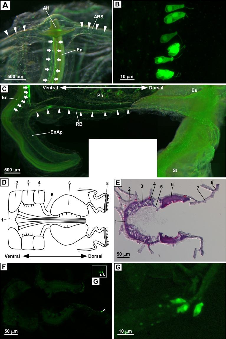
Figure 3.
Distribution of PC2 promoter-driven Kaede-positive cells in the endostyle. (A) Kaede-positive cells at the anterior end of the endostyle. Front view of the anterior end of the endostyle is shown. Arrows indicate Kaede-positive cells aligning in two rows in the endostyle. Arrowheads indicate Kaede-positive cells in the anterior pharyngeal blood sinus. The oral siphon was removed to make the endostyle more visible. Four images were merged to construct a deep-focus image. (B) Magnified image of the Kaede-positive cells in the endostyle. Kaede-positive ciliated cells in one row are shown. (C) Kaede-positive cells in the posterior end of the endostyle and pharynx. The posterior end of the endostyle and pharynx, and the endostylar appendix that extends out of the pharynx, are shown. Kaede-positive cells in the endostyle were observed continuously near the posterior end of the pharynx, and a small number of the cells was observed in the endostylar appendix (arrows). Arrowheads indicate Kaede signals along the blood sinus, which is also known as the retropharyngeal band. Fourteen images were merged to construct a composite image. (D) Schematic illustration of the horizontal section of the endostyle. Zone names are indicated by numbers. Short and long cilia are indicated by solid lines. (E) Hematoxylin-eosin staining of the horizontal section of the endostyle. Zone names are indicated by numbers. (F) Fluorescent image of the horizontal section of the endostyle. Kaede-positive cells in zone 7 are indicated by arrowheads. (G) Magnified image of the framed region in (F). Kaede-positive cells are localized in the dorsal terminal of zone 7, proximal to zone 8. The images in (A,C) were obtained under a fluorescence stereomicroscope as a superimposed image. The images in (B,F,G) were obtained under a confocal laser scanning microscope, and the image in (B) was obtained as a z-stack image. The image in (E) was obtained under a light microscope. ABS, anterior pharyngeal blood sinus; AH, anterior hood; En, endostyle; EnAp, endostylar appendix; Es, esophagus; Ph, pharynx; RB, retropharyngeal band; St, stomach.

