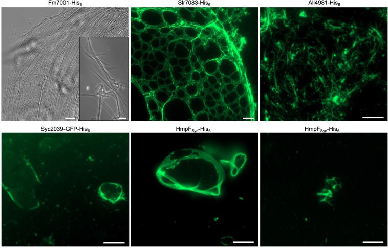
Figure 2.
Cyanobacterial CCRPs assemble into diverse filament-like structures in vitro. Bright field and epifluorescence micrographs of filament-like structures formed by purified and renatured Fm7001-His6 (0.7 mg ml−1), Slr7083-His6 (1 mg ml−1), All4981-His6 (0.5 mg ml−1), Syc2039-GFP-His6 (0.3 mg ml−1), HmpFSyc-His6 (0.5 mg ml−1) and HmpFSyn-His6 (0.5 mg ml−1). Proteins were dialyzed into 2 mM Tris-HCl, 4.5 M urea pH 7.5 (Fm7001), HLB (Slr7083), PLB (All4981, HmpFSyc, HmpFSyn) or BG11 (Syc2039). Renatured proteins were either directly analyzed by bright field microscopy (Fm7001) or stained with an excess of NHS-Fluorescein and analyzed by epifluorescence microscopy. The NHS-Fluorescein dye binds primary amines and is thus incompatible with urea, which is why Fm7001 filament-like structures were visualized by bright field microscopy. Scale bars: 10 µm or (Fm7001 inlay and Slr7083) 20 µm.

