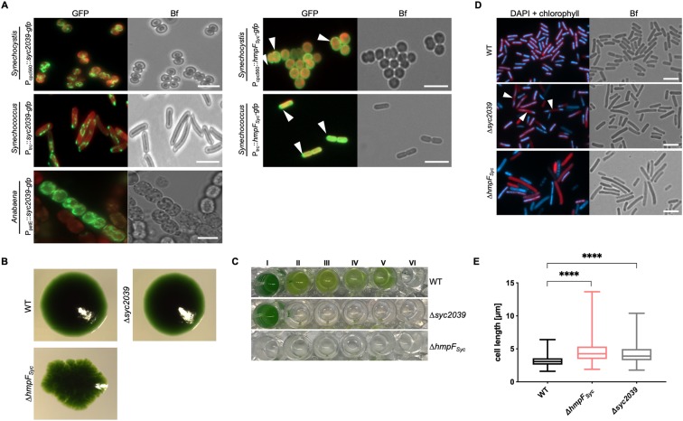
Figure 6.
Synechococcus CCRPs affect cytokinesis and cellular integrity. (A) Merged GFP fluorescence and chlorophyll autofluorescence (red) and bright field micrographs of Synechocystis, Synechococcus and Anabaena cells expressing Syc2039-GFP or HmpFSyc-GFP from Pcpc560, PpetE or Ptrc. Synechocystis cells were grown in BG11, Anabaena cells were grown in BG110 supplemented with 0.25 µM CuSO4 for 1 day, and Synechococcus cells were grown on BG11 plates supplemented with 0.01 mM (Syc2039) or 1 mM (HmpFSyc) IPTG. Micrographs of Synechococcus and Anabaena cells expressing Syc2039-GFP are maximum intensity projections of a Z-stack. White triangles indicate HmpFSyc-GFP spots. Attempts to translationally fuse a YFP-tag to the N-terminus of Syc2039 were unsuccessful, possibly due to the transmembrane domain predicted to the Syc2039 N-terminus (Supplementary Table 1). (B) Colony formation of Synechococcus WT and mutant strains on BG11 plates. (C) Cell viability of Synechococcus WT and mutant strains grown in (I) BG11 or BG11 supplemented with (II) 5 mM glucose, (III) 200 mM glucose, (IV) 2 mM NH4Cl, (V) 200 mM maltose or (VI) 500 mM NaCl. (D) Merged DAPI fluorescence and chlorophyll autofluorescence (red) and bright field micrographs of Synechococcus WT and mutant strains grown on BG11 plates and stained with 10 μg ml−1 DAPI. White triangles indicate non-dividing cells revealing inhomogeneous DNA placement. (E) Cell length of Synechococcus WT (n = 648), non-segregated ΔhmpFSyc (n = 417) and Δsyc2039 (n = 711) mutant cells. Values indicated with * are significantly different from the WT. ****P < 0.0001 (one-way ANOVA, using Turkey’s multiple comparison test). Scale bars: 5 µm.

