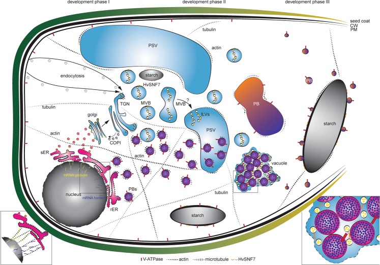
Figure 8.
Quantitative in situ mapping of the endomembrane system during barley endosperm development. Quantitative proteomics and in situ microscopic analyses identified HvSNF7 and MVBs as putative key players for protein sorting into PBs during barley starchy endosperm development. At developmental phase I, mRNA of e.g., globulin and e.g., hordein are transported by the cytoskeleton to the ER where they are entering different protein trafficking pathways (zoom in)59–63. During developmental phase I and II, PSVs become smaller and PBs are formed, both putatively regulated by the cytoskeleton. In parallel, MVBs containing HvSNF7 appear and possibly fuse with PSVs, leading to PSVs containing HvSNF7 positive ILVs and PBs (zoom in). Between developmental phase II and III, PSVs collapse, and PBs fuse to one big PB containing HvSNF7. At developmental phase III, PBs become smaller again, attaching to the protein matrix at the periphery of the starch granule. Note the additional localization of HvSNF7 at the starch granules between phase I and III. Additionally, V-ATPase localize to PBs at developmental phase I, acidifying PBs. V-ATPase could be further observed at starch granules during development. Schema is not in scale. PSV, protein storage vacuole; MVB, multivesicular body; ILVs, intraluminal vesicles; PB, protein body; sER, smooth ER; rER, rough ER; CW, cell wall; PM, plasma membrane.

