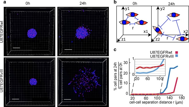
Fig. 1. U87EGFRvIII neurospheres demonstrate enhanced infiltrative properties in comparison with U87EGFRwt neurospheres.
a GBM neurospheres (NS) were embedded into 40% Matrigel (U87EGFRwt NS are shown in upper panel, and U87EGFRvIII NS in lower panel). Cell nuclei were imaged at 0 h (left panel) and 24 h (right panels) using confocal microscopy. Pink dots represent geometric centers of each nuclei which were used to define the cell coordinates. These coordinates were used to calculate cell–cell distances as described in Methods. Scale bars represent 150 μm. b Cell–cell separation distance (r) was calculated as described in Methods. All cell pairs, up to a separation distance of 200 μm were measured. c Fold change in the percentage of the cell pairs located at certain cell–cell distance, r, after 24 h relative to 0 h is presented in the plot. At least eight neurospheres were analyzed for each condition.

