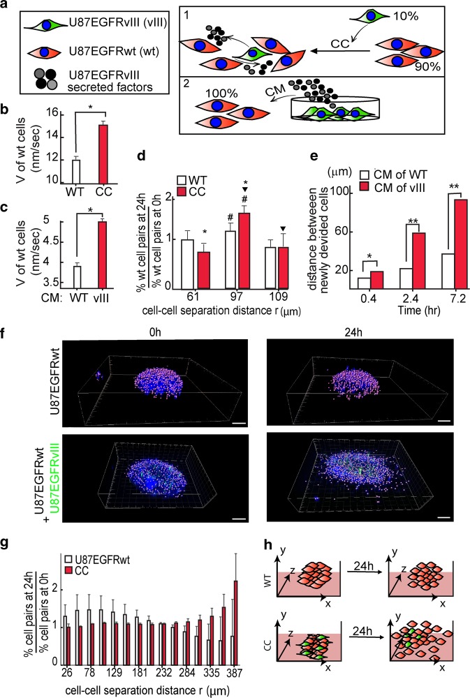
Fig. 2. U87EGFRvIII cells modulate the migration and spreading properties of U87EGFRwt cells.
a Effect of U87EGFRvIII cells (shown in green) on U87EGFRwt cells (shown in red) was examined by either co-culture (CC) or by stimulating U87EGFRwt cells with conditioned medium (CM) of U87EGFRvIII as shown in the illustration. b, c Change in the cell velocity (V) is shown for U87EGFRwt cells which were cultured for 24 h with either 10% of U87EGFRvIII cells (*P < 0.001) (b) or under CM of U87EGFRvIII cells (CM (vIII), *P < 0.001) (c). d U87EGFRwt cells were either cultured with 10% of U87EGFRvIII or alone. The fold change in the percentage of the cell pairs located at certain cell–cell distance, r, after 24 h relative to 0 h was calculated (the symbols #, ▾ indicate P < 0.05, the symbol * indicates P < 0.01). e U87EGFRwt cells were incubated with either U87EGFRvIII CM (vIII) or their own CM (WT) medium. U87EGFRwt dividing cells were imaged using live-cell NIS-Elements—Microscope Imaging. Cell–cell distances of dividing cells were measured every 20 min. Three representative time intervals are shown; n = 12 cell pairs were used for each condition (*P < 0.05; **P < 0.001). f U87EGFRwt neurospheres are shown in the upper panel and CC in the lower panel. Hoechst-labeled cell nuclei were imaged at 0 h (left panel) and 24 h (right panels) using confocal microscopy. U87EGFRwt cells were labeled with Qtracker 705, shown in green in the figure. Scale bars represent 100 μm. g The fold change in cell–cell separation distances were calculated after 24 h relative to 0 h. h The results in (a–g) suggest that U87EGFRvIII cells modulate the migration and spreading properties of U87EGFRwt through soluble factors secreted from U87EGFRvIII cells.

