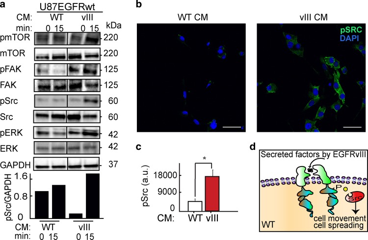
Fig. 4. Src mediates U87EGFRvIII/ U87EGFRwt crosstalk.
a Upper panel: Cells were grown under either U87EGFRwt conditioned medium (CM WT) or under U87EGFRvIII CM (CM vIII). Western blot assay was performed to examine the effect of U87EGFRvIII CM (vIII) on U87EGFRwt cells after 15 min. Bottom panel: quantification of pSrc levels normalized to GAPDH. All the proteins in the tested samples were assayed on the same membrane. The line in the middle indicates that non-relevant samples are not shown. b Representative images of immunofluorescence assays show Src activation (as represented by increase in pSrc) in U87EGFRwt cells following stimulation with CM of U87EGFRvIII (vIII). ×40 lens; scale bars represent 150 μm. c Average of pSrc intensity was quantified from eight fields (~20 cells/field). Plot is representative of at least three independent experiments, (*P = 0.005). Values are standard error (SE). d Schematic representation of Src activation in U87EGFRwt cells in response to EGFRVIII CM.

