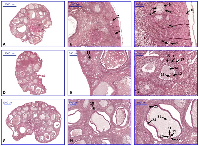
Figure 8.
Photomicrograph representing the sections of ovaries stained with hemotoxylin and eoesin. All images present follicles in various stages of development. The description of each arrow and number is represented in brackets. (A) Primordial and primary follicles. Tissue sections exhibiting: (B) Primordial follicles and primary follicles, and (C) primary oocytes, zona pellucida, granulosa cells, theca cells, follicular cells, cortical stroma and tunica albuginea. (D) Secondary follicle. Tissue sections exhibiting: (E) secondary follicles, and (F) primary oocytes, zona pellucida, antrum, granulosa cells, theca cells and theca externa. (G) Graafian follicle. Tissue sections exhibiting: (H) Graafian follicles, and (I) secondary oocytes, zona pellucida, corona radiata, cumulus oophorus, antrum, granulosa cells and theca externa. 1, primordial follicle; 2, primary follicle; 3, 7 and 12, primary oocyte; 4, 13 and 20, zona pellucida; 5, 15 and 24, granulosa cells; 6 and 16, theca cells; 8, follicular cells; 9, corticol stroma; 10, tunica albuginea; 11, secondary follicle; 14 and 23, antrum; 17 and 25, theca externa; 18, Graafian follicle; 19, secondary oocyte; 21, corona radiata; 22, cumulus oophorus.

