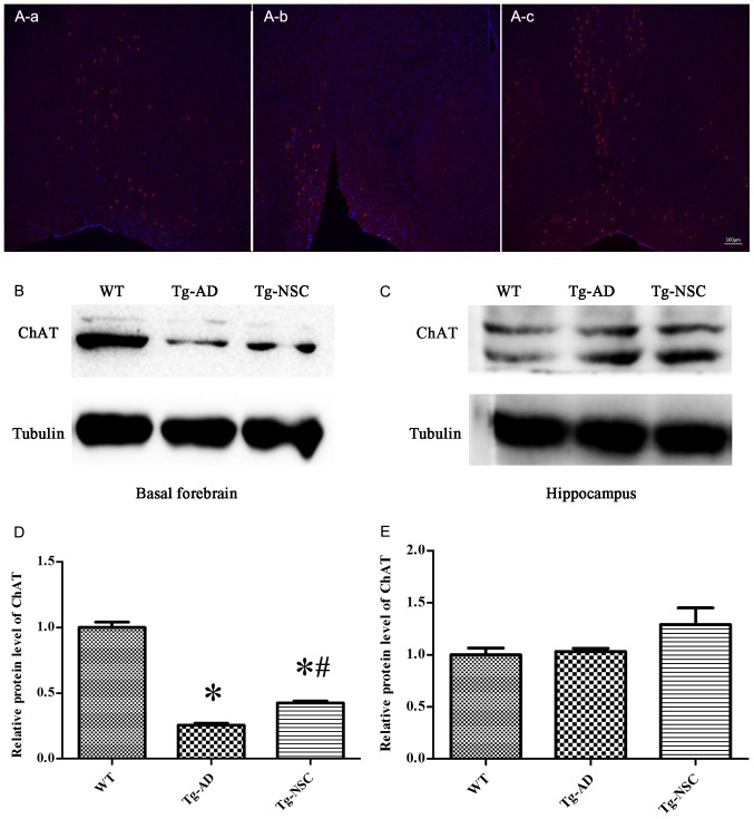
Figure 5.
Cholinergic neurons in the basal forebrain. (A) Confocal display the ChAT-positive neurons, (A-a) WT group; (A-b) Tg-AD group; (A-c) Tg-NSC group. (B and C) ChAT protein expression in the basal forebrain and hippocampus. (D and E) Relative levels of ChAT protein in the basal forebrain and hippocampus. *P<0.05 compared with WT group; #P<0.05 compared with Tg-AD group. ChAT, choline acetyltransferase; WT, wild-type; Tg, transgenic; AD, Alzheimer's disease; NSC, neural stem cell.

