Figure 4.
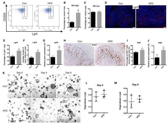
An HFD increases ISC count in the colon. Colon crypts were isolated, and epithelial cell suspensions were dissociated into single cells. (A) Representative flow cytometry analysis showing Lgr5 expression in the CD326 cell population obtained from the colon. (B) ISCs defined as Lgr5high, and (C) progenitor cells defined as Lgr5low in the entire colon. (D) Representative images of immunofluorescence staining for Lgr5, seen as red fluorescence, with DAPI used as a counterstain (scale bar, 50 µm; magnification, ×40). (E) Percentage of Lgr5+ cells among the crypt cells, and (F) the number of Lgr5+ cells per a crypt. (G) mRNA expression of Lgr5. (H) Representative images of immunohistochemical staining for Ki67 (scale bar, 100 µm; magnification, ×100), and (I) quantified Ki67+ cells as a percentage of the crypt cells. (J) mRNA expression of Ki67 in the colon. (K) Colon crypts were isolated from Con and HFD mice and cultured in Matrigel to develop organoid colonies. Representative images of colonic organoids at days 0, 4 and 6 are displayed (scale bar, 100 µm; magnification, ×20). Organoids per crypt at (L) day 4 and (M) day 6 are shown. Data are presented as the mean ± standard deviation. *P<0.05 and **P<0.01, vs. Con group. ISC, intestinal stem cell; HFD, high-fat diet; Con, control.

