Figure 5.
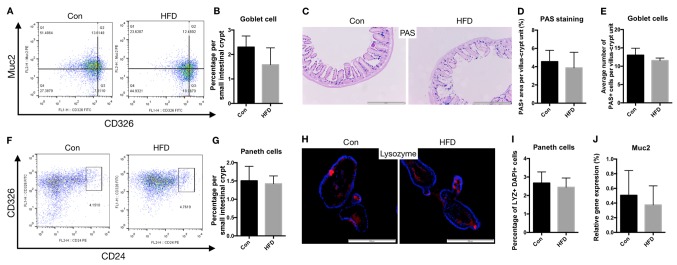
Barrier function of the small intestine was not affected by HFD. Goblet and Paneth cell numbers, and cell apoptosis were analyzed to determine the barrier function of the small intestine. (A) Representative flow cytometry analysis of Muc2 expression in CD326 cell populations. (B) Percentage of goblet cells in the small intestine. (C) Ileal sections were stained for goblet cells using PAS (scale bar, 500 µm; magnification, ×20). (D) Percentage of area stained as PAS-positive among the total villi-crypts sections. (E) Quantification of the number of goblet cells per villus-crypt unit. (F) Representative flow cytometry analysis showing CD24 expression in CD326 cell populations. (G) Percentage of Paneth cells in small intestinal crypts. (H) Crypt-derived organoids of Paneth cells, including lysozyme-positive cells, assessed using immunofluorescence (blue, DAPI; red, lysozyme; scale bar, 200 µm; magnification, ×40). (I) Quantification of Paneth cells in small intestinal crypt-derived organoids. (J) mRNA expression levels of Muc2 in the small intestine of the Con and HFD mice. HFD, high-fat diet; Con, control; PAS, Periodic Acid-Schiff; Muc2, mucin 2.

