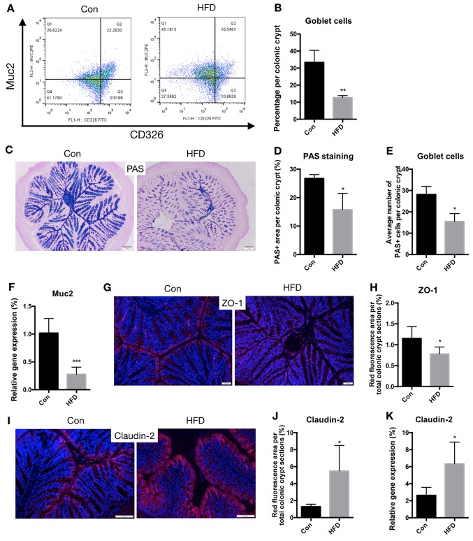
Figure 7.
An HFD compromises the barrier function of the colon. Goblet cell number, tight junctions and cell apoptosis were analyzed to determine the barrier function of the colon. (A) Representative flow cytometry analysis, showing Muc2 expression among the CD326 cell populations. (B) Proportion of goblet cells. (C) Histochemical PAS staining of goblet cells (scale bar, 100 µm; magnification, ×20), (D) percentage of area stained as PAS-positive among the colonic crypts, and (E) quantification of the average number of goblet cells per crypt. (F) mRNA expression of Muc2 in the colon. (G) Representative images of immunofluorescence staining for ZO-1 seen as red fluorescence, with DAPI used as the counterstain (scale bar, 50 µm; magnification, ×40), and (H) quantification of red fluorescence. (I) Representative images of immunofluorescence staining for Claudin-2 seen as red fluorescence, with DAPI used as the counterstain (scale bar, 100 µm; magnification, ×40), and (J) quantification of red fluorescence. (K) mRNA expression of Claudin-2 in the colon. *P<0.05, **P<0.01 and ***P<0.001, vs. Con group. HFD, high-fat diet; Con, control; PAS, Periodic Acid-Schiff; Muc2, mucin 2.

