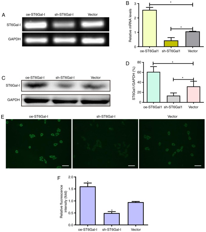
Figure 1.
Establishment of oe-ST6Gal-I and sh-ST6Gal-I subcell clones of OVCAR3 human ovarian cancer cells. (A and B) OVCAR3 cells were transfected with oe-ST6Gal-I, sh-ST6Gal-I and empty vector, and ST6Gal-I stable expression in clones was analyzed by reverse transcription-PCR. Protein levels of ST6Gal-I in oe-ST6Gal-I or sh-ST6Gal-I cells were assessed by (C) western blotting and (D) subsequent densitometry. *P<0.05. (E) α2,6-Linked sialic acid levels were detected with FITC-conjugated SNA lectin by fluorescence microscopy. Scale bar, 100 µm. (F) Relative fluorescence intensity of oe-ST6Gal-I or sh-ST6Gal-I cells. *P<0.05 vs. vector. oe, overexpression; ST6Gal-I, α2,6-sialyltransferase; sh, small hairpin.

