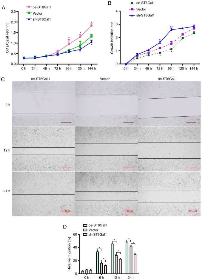
Figure 2.
Cells with high ST6Gal-I expression have enhanced viability and migratory ability. (A) Viability in oe-ST6Gal-I and sh-ST6Gal-I cells was measured by a CCK-8 assay. (B) Growth inhibition rates in serum-starved oe-ST6Gal-I and sh-ST6Gal-I cells were assessed by a CCK-8 assay. *P<0.05 vs. respective vector. (C) Scratch wound healing ability of oe-ST6Gal-I and sh-ST6Gal-I cells was determined by imaging cells under a microscope after incubation for 0, 12 and 24 h. (D) Quantitative analysis of the migratory ability of oe-ST6Gal-I and sh-ST6Gal-I cells. *P<0.05. ST6Gal-I; **P<0.01 vs. vector group/α2,6-sialyltransferase; oe, overexpression; sh, small hairpin; CCK-8, Cell Counting Kit-8; OD, optical density; Abs, absorbance.

