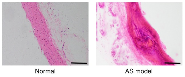
Figure 2.

Histological changes in the thoracic aortas of atherosclerotic rats induced by a high-fat diet identified by hematoxylin and eosin staining. In the normal group, the arterial intima was intact and smooth, and the cells were orderly arranged. In the AS model group, atherosclerotic plaque of the intima was present and the arrangement of the cells was disordered. Scale bars, 100 µm. AS, atherosclerosis.
