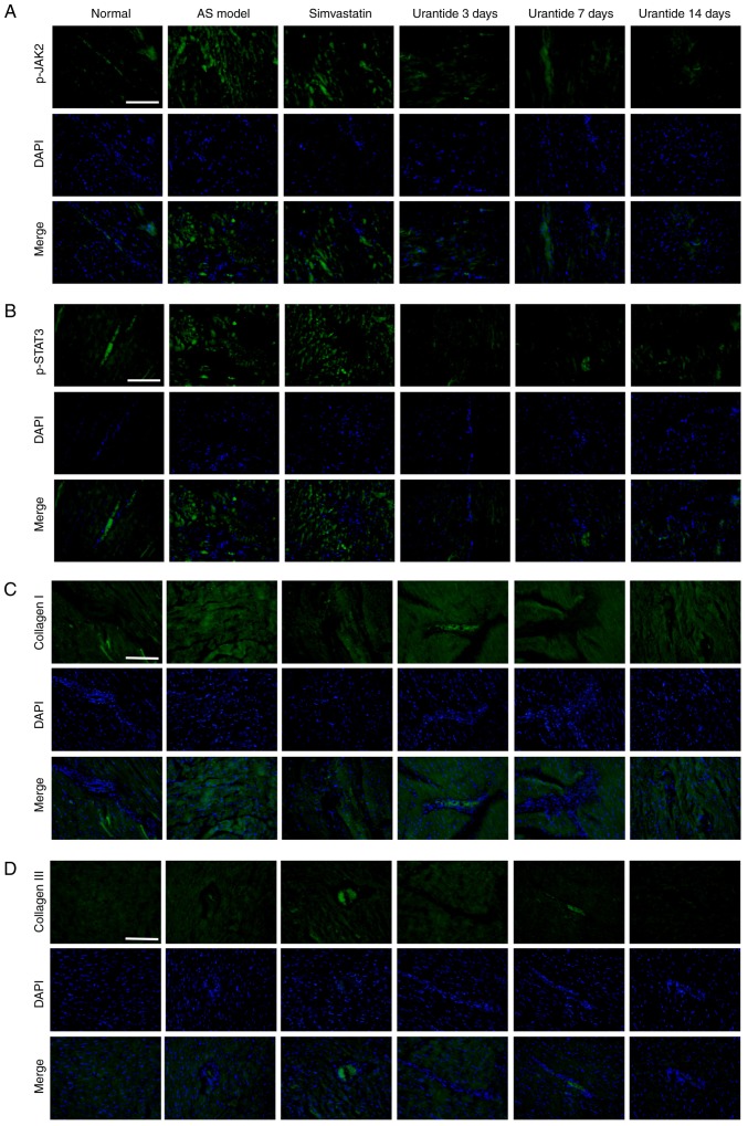
Figure 5.
Effect of urantide on the distribution and localization of p-JAK2/p-STAT3 and collagen type I/III. Immunofluorescence staining of: (A) p-JAK2, (B) p-STAT3, (C) collagen I and (D) collagen III indicating the location and intensity of the positive expression of these proteins in cardiac tissue derived from each group. Scale bars, 50 µm. Blue, nucleus; green, positive expression. AS, atherosclerosis; p-JAK2, phosphorylated-Janus kinase 2; p-STAT3, phosphorylated-signal transducer and activator of transcription 3.

