
Figure 2. Cerebral blood flow changes in rat brain.
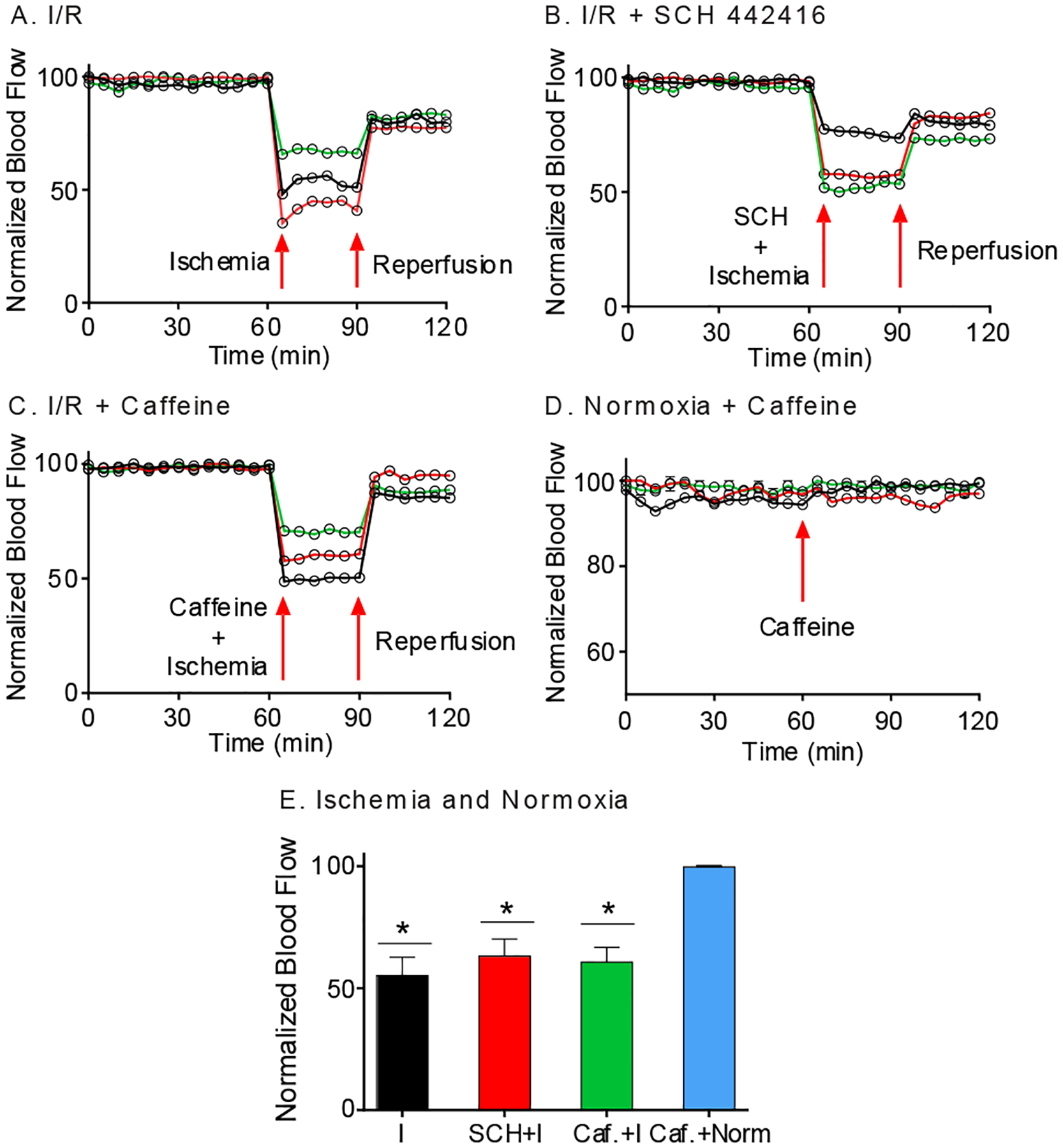
(A) Blood flow change during normoxia and I/R (n = 3 animals). Blood flow decreased during BCCAO and is restored, but not to baseline, during reperfusion. (B) Blood flow change during I/R with SCH 442416 (3 mg/kg) administration is similar to plot without drug (n = 3 animals). (C) Blood flow change during I/R with caffeine (100 mg/kg) administration is similar to that without caffeine. (D) Caffeine control (n = 3 animals). Blood flow was measured before and after caffeine (100 mg/kg) during normoxia. Blood flow did not change. (E) Average changes in blood flow. Blood flow significantly decreased during 30 min of ischemia (I) (*p = 0.027), SCH+ ischemia (*p = 0.034) and caffeine + ischemia (*p = 0.023) groups compared to 30 min of normoxia (Norm) Caffeine administration itself did not affect blood flow under normoxia (p = 0.94). Statistical analyses are paired t-test, where each treatment is compared with its own control.